Gutch Manish, DM
Senior Resident, Department of Endocrinology
D-15 Lala Lajpat Rai Memorial Medical College
Meerut-250004, Uttar Pradesh, India
Tel. No.: 453429252
E-mail: manish07gutch@gmail.com
e-ISSN 2308-118x
Printed in the Philippines
Copyright © 2015 by the JAFES
Received April 26, 2015. Accepted August 10, 2015.
Published online first: October 1, 2015.
Background. Celiac disease is frequently associated with type 1 diabetes mellitus, but is usually ill-defined and not usually suspected until the disease becomes advanced.
Objective. To study the prevalence and clinical profile of celiac disease among patients with type 1 diabetes mellitus in a tertiary care referral centre in north India.
Methodology. Two hundred and fifty six patients were screened (149 males and 107 females) during the study period of two years, patients were evaluated for the clinical signs, biochemical investigations and family history of celiac disease in tertiary care health center in western Uttar Pradesh.
Results.Twenty four (9.37%) patients were diagnosed to have celiac disease; the mean age at diagnosis of diabetes was 9.34 ± 7.3 years. Only 1/24 patients with celiac disease had been diagnosed before detection of diabetes mellitus. The common manifestations were normocytic normochromic anemia (66.6%) followed by diarrhoea (62.5%), abdominal pain/bloating sensation (58.3%) and short stature (58.3%). Some uncommon manifestations were also observed in small number of patients: rickets (20.8%), recurrent hypoglycemia (16.6%), carpopedal spasm (8.3%), and night blindness (8.3%).
Conclusion.Celiac disease was found in about 10% of patients with type 1 diabetes, almost 10-20 times higher than that observed in the general pediatric population. Atypical manifestations (rickets, recurrent hypoglycemia, carpopedal spasm and night blindness) were found to be common in patients with type 1 diabetes as compared to the general population. Unexplained anemia, diarrhoea, short stature and rickets should raise suspicion for the possibility of undiagnosed celiac disease in type 1 diabetes mellitus.
Keywords: type 1 diabetes mellitus, celiac diseases, short stature, anemia
Type 1 diabetes mellitus is a common autoimmune disorder of the pediatric population and it is frequently associated with other autoimmune conditions, especially with autoimmune hypothyroidism and celiac disease.1 Celiac disease (CD), or gluten-sensitive enteropathy, is an autoimmune disorder characterized by inflammation, villous atrophy and crypt hyperplasia of the small bowel mucosa after ingestion of dietary gluten and recovers when gluten-containing cereals are withdrawn from the diet.
The mean prevalence rate of celiac disease in type 1 diabetes patients varies in studies but ranges from 1% to 11%,2 almost 10-20 times higher than that observed in general pediatric population.3 It has an incidence of 1 in 96 in north India.4
The presentation of celiac disease in Type 1 diabetes patients is extremely variable, less than one third of patients present with gastrointestinal complaints, and some patients remain asymptomatic and are only diagnosed during routine screening procedures.5 The prominent extraintestinal manifestations of celiac disease are short stature, delayed puberty, poor glycemic control, nutritional anemia, etc.6-7
Patients with celiac disease frequently present with growth failure, malnutrition, hypocalcemia anemia, suboptimal growth velocity and poor weight gain and pathogenesis appears to be multifactorial A few studies support the role of RANKL/OPG system in the pathogenesis, but the exact cause for it is still unknown.8
The main aim of this cross-sectional analytic study is to study the prevalence and clinical characteristics of celiac disease in patients with type 1 diabetes mellitus in Northern India.
Two hundred and thirty six children and adolescents with type 1diabetes, aged 6 to 18 years, presenting to the endocrine OPD or admitted to the endocrinology ward, were enrolled in the study period of two years from July 2011 to June 2013. Ethical clearance for the study was secured from the institution
After explaining the objectives of the study, a written informed consent was obtained from the patients or their parents. Patients were evaluated clinically, biochemically and inquiries were made for family history of other common autoimmune disorders, which are associated with type 1 diabetes.
Physical examination along with Tanner scoring was done by two endocrinologists, which included one pediatric endocrinologist. Blood samples were collected for: anti-TTG immunoglobulin subclass A (IgA) using enzyme-linked immunosorbent assay (ELISA), CBC, iron profile, glycosylated hemoglobin (HbA1C), calcium, phosphorus and albumin.
X-rays of the wrist and knee were done in all patients of celiac disease in the hospital premises and the x-rays were reported first by an experienced endocrinologist and then by radiologists, and if both agreed on a finding, it was accepted. Bone age was assessed using the Tanner Whitehouse 2 system.
Delayed bone age was defined as difference of at least 24 months between chronological age and bone age. Vitamin D level was done when appropriate.
Endoscopic duodenal biopsies were undertaken for those who were negative for anti-TTG antibody after informed consent. Screening for other autoimmune disorders was done only when signs or symptoms or family history suggestive of the disorder were present.
Statistical AnalysisAll categorical variables were expressed as percentages and all continuous variables were expressed as mean ± standard deviation. Categorical variables were compared using Fisher's exact test and Chi-square test, whichever is applicable. Continuous variables were compared using independent t-test and ANOVA as applicable. All p values <0.05 were taken as significant. Bivariate correlations were calculated using Pearson's correlation coefficient. Statistical analysis was performed by using software SPSS version 17.
Table 1 shows the comparison between the patients with and those without celiac disease, the age of presentation with celiac disease was almost one year later than the age of presentation of patients without celiac disease.
Click here to download Table 1Table 1. Demographic and clinical characteristics of patients
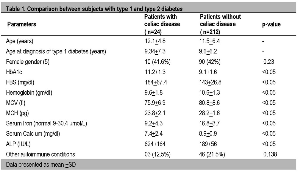
Patients with celiac disease had much higher HbA1C, FBS and ALP in comparison with patients without celiac disease, which both are significant (<0.05), but had lower hemoglobin, MCV, MCH, serum iron, and serum calcium and all were significant (<0.05). Sex ratio was found to be equal in both, while the incidence of other autoimmune conditions was found to be non- significant.
Table 2 shows the demographic profile of patients with celiac disease and the most common manifestations
Click here to download Table 2Table 2. Clinical profile of children with type 1 diabetes associated with celiac disease, N=24, Uttar Pradesh, India
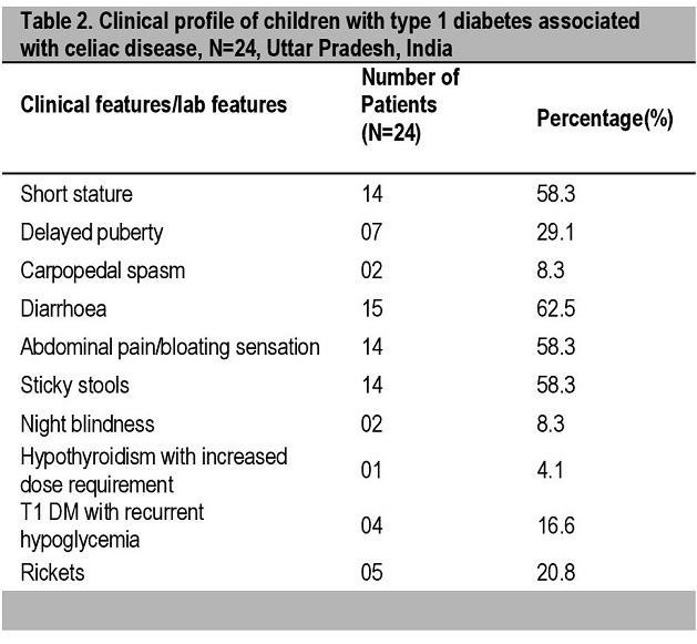
Table 3 shows the prevalence of different laboratory and biochemical abnormalities in the T1DM patients with celiac disease. The data shows that anemia was the most common laboratory finding with an overall prevalence rate of 87.4% (66.6% normocytic normochromic and 20.8% microcytic hypochromic).
Click here to download Table 3Table 3. Laboratory results/manifestation of celiac disease
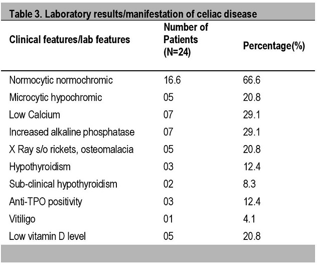
In bone and calcium metabolism abnormalities, the prevalence of both low calcium and increased alkaline phosphatase was 29.1% while 20.8% of patients had radiological signs of rickets/osteomalacia and hypovitaminosis D. Some 12.4% of patients suffered from overt hypothyroidism with the number of individuals having Anti-TPO positivity but only 8.3% of patients had subclinical hypothyroidism.
This is a cross-sectional study carried out in the department of endocrinology and metabolism during the study period of from July 2011 to June 2013. The main aim of the study was to determine the prevalence and various clinical manifestations of celiac disease in type 1 diabetes mellitus.
Celiac disease is an autoimmune-mediated enteropathy precipitated by the ingestion of gluten-containing foods (including wheat, rye and barley) in genetically susceptible persons. Celiac disease is also frequently associated with type 1 diabetes mellitus but less frequently than autoimmune thyroiditis. From last few years, numerous screening studies showed increased worldwide prevalence of celiac diseases in type 1 diabetes mellitus5 However, data from South Asia is very limited especially in the Indian subcontinent.9 Its prevalence in children and adolescents with type 1 diabetes ranges from 5 to 7%. In a previous study done by Bhadada et al10 the prevalence rate was 11.1% among type 1 diabetics. In the present study, out of 236 type 1 diabetics, celiac disease was present in 24 patients, the prevalence is 9.37%. Only one patient was diagnosed with celiac disease before the diagnosis of type 1 diabetes.
Gluten intake varies from population to population and depends upon dietary practices. Wheat is the staple cereal in the northern part of India and flat bread made from wheat flour is one of the most important constituents of almost every meal. In the southern and northeastern part of India, rice is a staple food. A typical North Indian diet, where flat bread is the usual meal, contains about 25-30 g of gluten per day; whereas average gluten intake in the West varies from 10 to 20 g/day.23
Although the majority of detected cases of celiac diseases in children with diabetes mellitus are reported to be asymptomatic or silent,12,13 we have demonstrated that many children do have subtle gastrointestinal complaints that may indicate celiac diseases. Directing specific questions may help increase the yield of gastrointestinal symptoms in these children.
In our study, the common clinical manifestations which were found in type 1 diabetes mellitus with celiac disease are diarrhoea (62.5%), abdominal pain/bloating sensation (58.3%), short stature (58.3%), sticky stool (58.3%) and delayed puberty in 29.1% of patients. Some uncommon manifestations are rickets (20.8%), recurrent hypoglycemia (16.6%), carpopedal spasm (8.3%) and night blindness (8.3%). The most common laboratory findings were normocytic normochromic anemia (66.6%) followed by microcytic hypochromic anemia.
A concern with malabsorptive disease is that it could increase the incidence of hypoglycemia in diabetes, particularly in patients under tight control.21 About 16% patients presents with hypoglycemia.21
Malabsorption may also be linked to vitamin A deficiency which leads to night blindness and treatment failures may be due to inadequate management either of celiac disease or of thyroid disease or both, which may lead to increased dose requirement of levothyroxine.22
Numerous studies from all over the world showed the varied manifestations of celiac disease in type 1 diabetes, but these all depend upon the place of study, study population, follow up rate, etc. The most common manifestations in studies is the anemia followed by abdominal complains, short stature, delayed puberty, recurrent hypoglycemia and hypocalcemia.11,12
Studies on the impact of celiac diseases on glycemic control and growth in patients with T1D have shown conflicting results.13-14 Our patients with type 1 diabetes and celiac disease had poor glycemic control and growth parameters as compared to patients without celiac disease as judged by fasting blood sugar and HbA1C.
We confirmed the observation that children with diabetes who went on to develop celiac disease were younger at diagnosis of DM than other children with DM.13 The predominance of males with celiac disease in the present study has been observed in few studies,17 while other studies in different races have shown a female predominance,16 which likely represents variability of genetic and environmental factors among different races.
While the majority of our patients with celiac diseases were found to have gastrointestinal symptoms, iron deficiency and hypocalcemia, which are indices of malabsorption, were also prominent in the celiac disease patients15 The potential for early reversal of abnormalities in indices of intestinal malabsorption (iron and calcium deficiencies) is one of the advantages for screening asymptomatic children for early detection of celiac diseases in T1DM patients.
There are many barriers to maintenance of a gluten-free diet (GFD); some of them are universal and some of them are unique to Indian patients with CD. Patients with CD are challenged with barriers in maintenance of a strict GFD primarily due to inadequate information and education about the disease.23 Insufficient background awareness about CD and its strict dietary restriction in the community creates a problem for the patient and the family of patients with CD. Due to the lack of gluten labeling on food items in India, it is difficult for anyone to know if a particular food product is gluten-free or not. Contamination of food with gluten is another concern.23
Adherence to GFD is the most critical factor for remission of CD. Adherence to GFD is complex and is influenced by knowledge, country or region of residence, availability of GF food, determination, and social support. Gluten-containing foods can be replaced by rice, maize, barley grain, millet (Bajra), sorghum (Jowar) and other locally available millet flour or products.23 Following the introduction of gluten-free diet to patients, a sense of general well-being pervades, weight and height improve and the severity of acute and chronic complications of celiac disease decreases.22
Celiac disease was found in about 10% of patients with type 1 diabetes, almost 10-20 times higher than that observed in general pediatric population. Atypical manifestations (rickets, recurrent hypoglycemia, carpopedal spasm, and night blindness) were found to be common in patients with type 1 diabetes as compared to the general population. Unexplained anemia, poor glycemic control, diarrhoea, short stature and rickets should raise suspicion for the possibility of undiagnosed celiac disease in patients with type 1 diabetes mellitus. Growth indices and parameters of glycemic control improve considerably on timely detection and management of celiac disease.
1. Cappa M, Bizzarri C, Crea F. Autoimmune thyroid diseases in children. J Thyroid Res. 2010;2011(2011):675703. http://dx.doi.org/10.4061/2011/675703.
2. Kakleas K, Karayianni C, Critselis E, Papathanasiou A, Petrou V, Fotinou A, et al. The prevalence and risk factors for coeliac disease among children and adolescents with type 1 diabetes mellitus. Diab Res Clin Pract. 2010;90(2):202-208. http://dx.doi.org/10.1016/j.diabres.2010.08.005. 3. Mäki M, Mustalah K, Kokkonen J, Kulmala P, Haapalahti M, Karttunen T, et al. Prevalence of celiac disease among children in Finland. N Engl J Med. 2003;348:2517-2524. http://dx.doi.org/10.1056/NEJMoa021687. 4. Makharia GK, Verma AK, Amarchand R, Bhatnagar S, Das P, Goswami A, et al. Prevalence of celiac disease in the northern part of India: A community based study. J Gastroenterol Hepatol. 2011;26:894-900. 5. Gujral N, Freeman HJ, Thomson ABR. Celiac disease: Prevalence, diagnosis, pathogenesis and treatment. World J Gastroenterol. 2012;18(42):6036-6059. http://dx.doi.org/10.3748/wjg.v18.i42.6036. 6. Rojas-Villarraga A, Amaya-Amaya J, Rodriguez-Rodriguez A, Mantilla RD, Anaya JM. Introducing polyautoimmunity: Secondary autoimmune diseases no longer exist. Autoimmune Dis. 2012;2012(2012):254319.http://dx.doi.org/10.1155/2012/254319. 7. Rostami Nejad M, Rostami K, Pourhoseingholi MA, et al. Atypical presentation is dominant and typical for coeliac disease. J Gastrointestin Liver Dis. 2009;18:285-91. 8. Galluzzi F, Stagi S, Salti R, et al. Osteoprotegerin serum levels in children with type 1 diabetes: A potential modulating role in bone status. Eur J Endocrinol. 2005;153:879-885. http://dx.doi.org/10.1530/eje.1.02052. 9. Cerruti F, Bruno G, Chiarelli F, Lorini R, Meschi F, Sacchetti C and the Diabetes Study Group of the Italian Society of Pediatric Endocrinology and Diabetology. Younger age at onset and sex predict celiac disease in children and adolescents with type 1 diabetes: An Italian multicenter study. Diabetes Care. 2004;27(6):1294-1298. http://dx.doi.org/10.2337/diacare.27.6.1294. 10. Bhadada SK, Kochhar R, Bhansali A, Dutta U, Kumar PR, Poornachandra KS, et al. Prevalence and clinical profile of celiac disease in type 1 diabetes mellitus in north India. J Gastroenterol Hepatol. 2011;26(2):378-381. http://dx.doi.org/10.1111/j.1440-1746.2010.06508.x. 11. Goh C, Banerjee K. Prevalence of coeliac disease in children and adolescents with type 1 diabetes mellitus in a clinic based population. Postgrad Med J. 2007;83:132-6. http://dx.doi.org/10.1136/pgmj.2006.049189. 12. Bashiri H, Keshavarz A, Madani H, et al. Celiac disease in type I diabetes mellitus: Coexisting phenomenon. J Res Med Sci. 2011;16:401-6. 13. Amin R, Murphy N, Edge J, Ahmed ML, Acerini CL, Dunger DB. A longitudinal study of the effects of a gluten-free diet on glycemic control and weight gain in subjects with type 1 diabetes and celiac disease. Diabetes Care. 2002;25(7):1117-1122. http://dx.doi.org/10.2337/diacare.25.7.1117. 14. Kaspers S, Kordonouri O, Schober E, Krause U, Schimmel U, Hauffa BP, et al. Anthropometric parameters, metabolic control and thyroid autoimmunity in 127 biopsy-positive patients with type 1 diabetes and coeliac disease (CD) compared to 18,470 diabetic subjects without CD. 18th Congress of the International Diabetes Federation, Paris, 24-29. Diabetologia. 2003;46(S2):A232-3(presentation 671). 15. Buysschaert M, Tomasi JP, Hermans MP. Prospective screening for biopsy proven coeliac disease, autoimmunity and malabsorption markers in Belgian subjects with type 1 diabetes. Diabet Med. 2005;22(7):889-892. http://dx.doi.org/10.1111/j.1464-5491.2005.01542.x. 16. Shahbazkhani B, Faezi T, Akbari MR, Mohamadnejad M, Sotoudeh M, Rajab A, et al. Coeliac disease in Iranian type I diabetic patients. Dig Liver Dis. 2004;36(3):191-194. http://dx.doi.org/10.1016/j.dld.2003.10.015. 17. Araújo J, da Silva GAP, de Melo FM. Serum prevalence of celiac disease in children and adolescents with type 1 diabetes mellitus. J Pediatr (Rio J). 2006;82(3):210-214. http://dx.doi.org/10.2223/JPED.1478. 18. Goh VL, Estrada DE, Lerer T, Balarezo F, Sylvester FA. Effect of gluten-free diet on growth and glycemic control in children with type 1 diabetes and asymptomatic celiac disease. J Pediatr Endocrinol Metab. 2010;23(11):1169-1173. http://dx.doi.org/10.1515/jpem.2010.183. 19. Hansen D, Brock-Jacobsen B, Lund E, Bjørn C, Hansen LP, Nielsen C, et al. Clinical benefit of a gluten-free diet in type 1 diabetic children with screening-detected celiac disease. Diabetes Care. 2006;29(11):2452-2456. http://dx.doi.org/10.2337/dc06-0990. 20. Valerio G, Spadaro R, Iafusc, D, Lombardi F, del Puente A, Esposito A, et al. The influence of gluten free diet on quantitative ultrasound of proximal phalanxes in children and adolescents with type 1 diabetes mellitus and celiac disease. Bone. 2008;43(2):322-326. http://dx.doi.org/10.1016/j.bone.2008.04.004. 21. Schwarzenberg SJ, Brunzell C. Type 1 diabetes and celiac disease. Overview and medical nutrition therapy. Diabetes Spectrum. 2002;15(3):197-201. http://dx.doi.org/10.2337/diaspect. 15.3.1.97. 22. Collin P, Kaukinen K, Välimäki M, Salmi J. Endocrinological disorders and celiac disease. Endocr Rev. 2002;23(4):464-483. http://dx.doi.org/10.1210/er.2001-0035. 23. Price S. Understanding the importance to health of a balanced diet. Nurs. Times. 2005;101(1): 30-31.Authors are required to accomplish, sign and submit scanned copies of the JAFES Declaration that the article represents original material that is not being considered for publication or has not been published or accepted for publication elsewhere.
Consent forms, as appropriate, have been secured for the publication of information about patients; otherwise, authors declared that all means have been exhausted for securing such consent.
The authors have signed disclosures that there are no financial or other relationships that might lead to a conflict of interest. All authors are required to submit Authorship Certifications that the manuscript has been read and approved by all authors, and that the requirements for authorship have been met by each author.